Introduction
In the realm of life science publications, image integrity is a cornerstone of credibility and authenticity. With the advent of digital image-manipulation tools, ensuring the integrity of images used in scientific research has become both a significant challenge and a necessity.
This post delves into the importance of image integrity, the issues faced in maintaining it, and the solutions available to tackle these challenges.
The Importance of Image Integrity in Life Science Publications
Images, ranging from simple graphs to complex microscopy images, play a pivotal role in life science research. They provide a visual representation of data, making complex information easier to understand. Consequently, the integrity of these images is paramount. Any manipulation that leads to misrepresentation of the original data can lead to incorrect conclusions and, in severe cases, scientific misconduct.

The Challenges of Image Manipulation
With the increasing use of digital tools for image acquisition and processing, the risk of image manipulation, both intentional and unintentional, has escalated. Researchers may make minor alterations to images for various reasons, such as enhancing contrast or color balance to emphasize a particular point. However, these alterations can sometimes misrepresent the original data, leading to ethical issues.
Unacceptable Image Adjustments
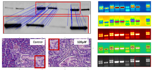
While basic adjustments applicable to all images within a figure, such as linear contrast and brightness, are generally permissible, other alterations like selective enhancements, deletions, or additions of elements within images are considered unacceptable. This category includes cut and paste in Western blotting, bands’ deletion, cloning within any images, etc. Such manipulations can significantly distort the data being represented and may lead to false interpretations.
The Role of AI in Ensuring Image Integrity
In response to these challenges, AI-powered solutions like Proofig are revolutionizing the way image integrity is maintained in scientific publications. Proofig offers automated image proofing for scientific research, ensuring that the images used adhere to ethical guidelines and are free from unacceptable manipulations. Its AI is capable of detecting any form of duplication, helps identify cut-and-paste artifacts, and can spot bands’ deletion in Western blots. This not only helps in preserving the credibility of the research but also significantly reduces the time and effort involved in manual image checking.

Conclusion
Maintaining image integrity in life science publications is crucial for the credibility and authenticity of scientific research. While the digital age has brought with it challenges in the form of easy image manipulation, it also provides innovative AI-powered solutions like Proofig’s AI tool to tackle these issues. As researchers, publishers, and academic institutions, it is our collective responsibility to uphold the highest standards of integrity in scientific publishing.
Tip
Always keep a record of the original images and the settings used for image acquisition and processing. This can be extremely useful if questions about the image’s authenticity arise in the future.
Frequently asked questions
Why does image integrity matter so much in life science research?
Because figures carry the evidence, and a manipulated or duplicated image can lead to incorrect conclusions or, in severe cases, misconduct findings. Life science papers depend on images, from microscopy and Western blots to graphs. Proofig AI, the image integrity platform trusted by the Science family of journals, screens every figure for duplication and reuse, manipulation, image plagiarism and AI-generated images, so authors, publishers and institutions catch problems before publication.
How does AI help keep life science images trustworthy?
AI checks every figure consistently and in minutes, catching duplications and edits that manual checks by eye can miss. Proofig AI pioneered automated pre-publication image integrity screening. Proofig AI compares each image within the manuscript and against the industry’s largest image-plagiarism database, with more than 155 million PubMed Open Source Database images, and flags manipulation and AI-generated images. Every finding is flagged for your review, and authors and editors make the final call.
Do I still need my original image files after screening figures with Proofig AI?
Yes, keep them, because original files let you check any region Proofig AI flags against the raw data. Proofig AI surfaces suspected issues for human review and does not make final scientific judgments, so your own records help settle each finding. Keeping raw images together with the acquisition and processing settings you used also protects you if a journal, reader or integrity office questions a figure after publication.
Why has image manipulation become a bigger risk in life science research?
Digital acquisition and editing tools make it easy to alter an image, intentionally or not, and generative AI can now create convincing fakes. Selective enhancements, deletions and added elements can distort results even when made innocently. Proofig AI detects cloning, splicing, deletion and content-aware edits within each sub-image, flags AI-generated images, and keeps updating its detection models as new manipulation methods and AI risks emerge.