

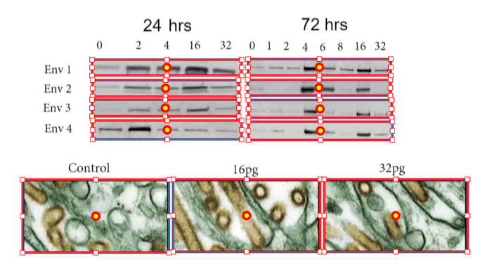
The world of life science research is teeming with diverse types of images, each carrying unique and valuable information. This blog post delves into the technology behind life science image detection, providing a comprehensive overview of the process.
The Challenge of Image Detection
Detecting images in a scientific manuscript is the first step in the process, and this is where image detection software excels—distinguishing between various visual elements and extracting only the scientific images. This involves distinguishing images from the surrounding text and other elements of the manuscript. While this may seem straightforward, this is no simple task for software, as images can vary greatly in size, shape, and complexity and may be embedded in different parts of a document.
The Power of AI in Image Extraction: How Image Detection Software Transforms Research
Artificial intelligence (AI) technology has revolutionized the process of image extraction. AI algorithms can scan a document and accurately identify and extract all images, regardless of their size or location in the manuscript. This technology is not only highly accurate but also incredibly fast, capable of analyzing entire papers in minutes.
Classifying Images Through Image Detection Software: A Step Further
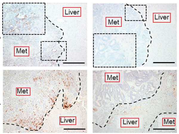
Once the images have been extracted, the next step entails classifying them by type. This is where the real challenge begins. Life science research utilizes a wide range of image types, including microscopy images, western blots, flow cytometry images, and more. Each of these image types carries different information and requires a different approach for analysis.
Text Overlay Detection: An Added Complexity
In addition to detecting and classifying images, the AI system also needs to scan each image for overlaying text. Text overlays are common in scientific images and are often used to label various parts of the image or provide additional information. Detecting and ignoring these text overlays is crucial, as they can significantly interfere with the image analysis.
The Importance of Accuracy
The process of life science image detection and classification needs to be highly accurate. Even small errors can lead to significant issues, such as misinterpretation of data and incorrect conclusions. Therefore, the AI algorithms and image detection software used for this process are designed to be as precise as possible so as to minimize the risk of errors.
Conclusion
The technology behind life science image detection is complex and sophisticated, but it plays a crucial role in maintaining the integrity of scientific publications. By accurately detecting and classifying images, image detection software helps ensure that the information presented in scientific manuscripts is reliable and accurate.
Tip
When preparing your manuscript for publication, consider using an AI-powered tool like Proofig AI to check the integrity of your images. This can save you time and help avoid potential issues down the line.
Frequently asked questions
What problems does Proofig AI’s image detection software find in a manuscript’s figures?
Proofig AI finds duplication and reuse, manipulation, image plagiarism and AI-generated images in every figure of an uploaded manuscript, with results in minutes. Proofig AI screens down to each sub-image and compares visuals with the industry’s largest image-plagiarism database, with more than 155 million PubMed Open Source Database images. Every finding is flagged for your review, using forensic tools and the Review Assistant, and only the findings the reviewer chooses go into the exportable PDF report.
Do I need to split multi-panel figures into separate images before uploading to Proofig AI?
No: upload the whole article to Proofig AI as one PDF or Word file, and it analyzes every figure automatically, with no formatting changes. Proofig AI works at the level of sub-images, the distinct visual elements inside a figure, such as one microscopy image, one line of Western blot bands or one graph. A single submission can hold up to 750 sub-images, in a file of up to 50 MB and 200 pages, with higher limits for enterprise accounts.
Does Proofig AI check each panel on its own or look across the whole manuscript?
Both: Proofig AI looks across every visual in a manuscript for duplication and reuse, and inside each sub-image for manipulation. The cross-figure check catches copies that were resized, rotated, flipped, cropped or partially overlapped. The manipulation check looks for cloning, splicing, deletion and content-aware edits. Manuscripts uploaded together are analyzed separately, each with its own report, and are not compared with each other. Every flag is a signal for human review, not a verdict.
Can image detection software review the rest of my paper, not only the figures?
Yes, through Octym: Proofig AI checks the figures, and Octym, from the same team, reviews the entire paper with 26 checks. Octym runs Proofig AI’s image checks plus checks on references, statistics, methods and metadata, all in one report. Instead of separate uploads and separate reports, or asking a general-purpose AI chatbot that runs no forensic pixel comparison across panels, authors get image forensics and a full manuscript review from the team behind Proofig AI. RefGuard, Proofig AI’s reference integrity product, flags retracted papers, hallucinated references and unsupported claims.