Proofig AI for Ethics Committees: Uphold the Highest Standards of Research Oversight
Automated image-integrity screening that supports ethical review, safeguards participant trust, and streamlines protocol evaluations.
Why Image Integrity Matters
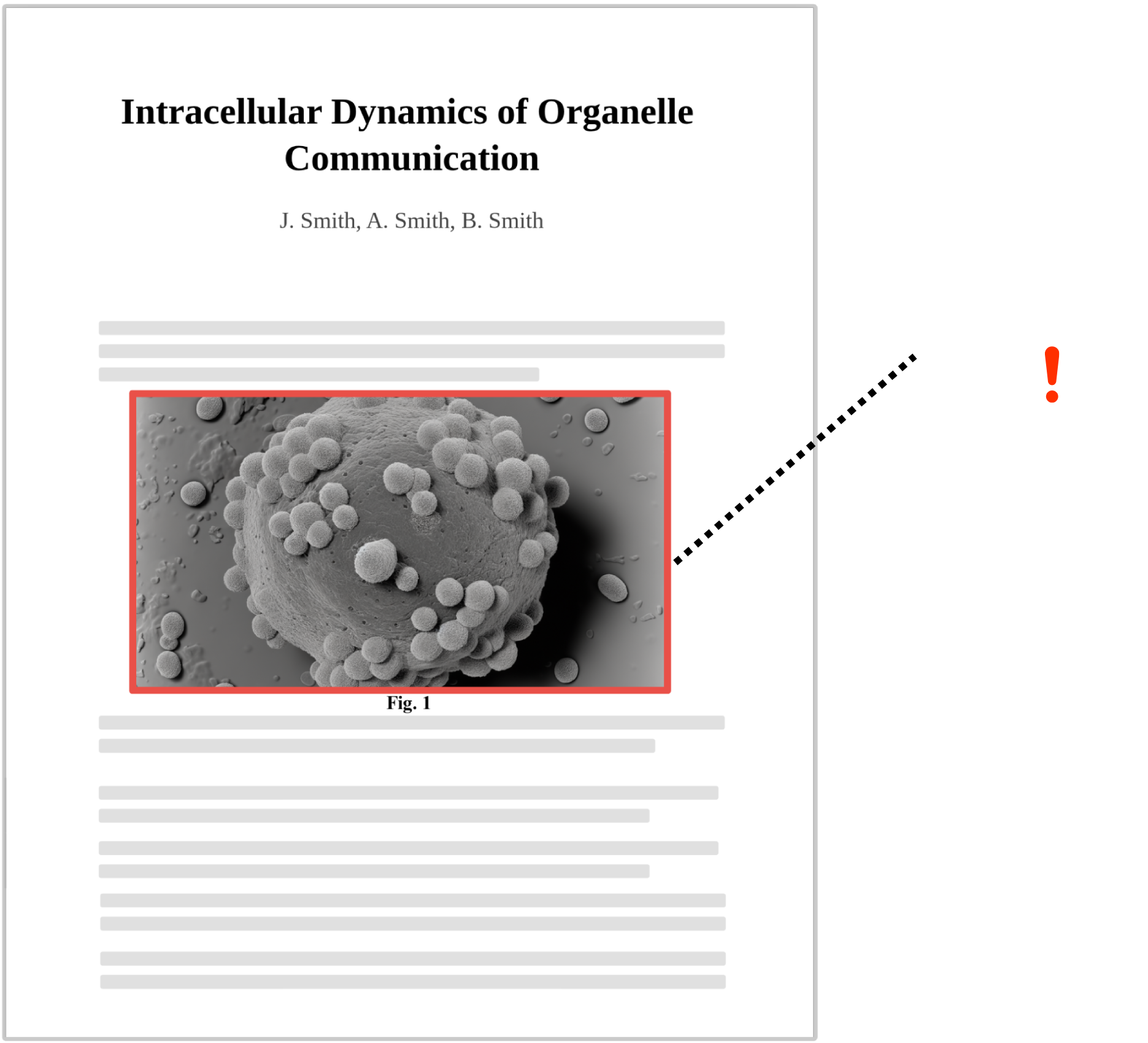
Roughly one-third of life-science manuscripts contain image issues—many discovered only after submission. Manipulated or duplicated figures can compromise study validity, prompt ethics investigations, and undermine public confidence in research.
Ethics committees must verify that study materials meet rigorous quality standards, yet manual review often misses subtle or large-scale problems. Automated screening delivers consistent, timely detection—helping boards flag concerns early and maintain robust oversight across every protocol.
Misconduct
Risk
Undetected image issues can invalidate findings and trigger formal inquiries.
Public & Participant
Trust
Integrity lapses erode confidence in approved studies and institutional review.
Limited Review
Time
Volunteer committees and small offices can’t inspect every figure by eye.
Why Ethics Committees Choose Proofig AI
Research institutions cannot directly oversee researchers’ manuscripts and images prior to submission, yet post-publication image issues directly harm institutional credibility—making automated image integrity solutions essential for proactive protection.
Wide-Ranging
Detection
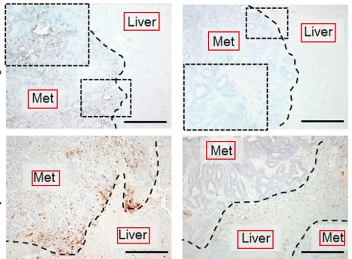
Identify duplication, manipulation, AI-generated content, and more within minutes.
Audit-Ready
Reporting
Download clear PDF reports to document findings for board records or regulators.
Secure &
Confidential
All files are processed on private servers; data is never reused for other purposes.
Flexible
Workflow
Upload protocol PDFs manually or integrate Proofig with your existing submission portal.
Dashboard
Oversight
Track screening activity, detection results, and remediation steps across studies.
Dedicated
Support
A named success manager assists with onboarding, training, and policy alignment.
How It Works
Choose Your Workflow Drag-and-drop a protocol PDF or integrate Proofig for automatic screening.
AI-Powered Analysis The system scans all images and sub-images for potential integrity issues.
Review and Validate Check flagged images, investigate further, and decide what to include in the committee report.
Download a Final Report Generate a detailed PDF to archive with meeting minutes, compliance records, or investigator feedback.
Trusted by Leading Institutions and Publishers


I believe Proofig is a very valuable tool for duplication detection, and suited to effectively assist journals’ image integrity screening routines.
Further Resources
- Proofig AI and MDPI Sign Multi-Year Deal to Strengthen Research Integrity
- AI-Generated Image Detection: A Leap in Scientific Integrity
- Science Journals Adopt AI-Powered Image Integrity Checks